

A routine dental exam does far more than tidy up your smile and polish teeth for a brighter look. The visit gives clinicians a chance to scan for subtle signs that point to early trouble and to map out what comes next if an issue shows up.
Regular checks create a rhythm of care that keeps small matters small and prevents them from ballooning into bigger headaches.
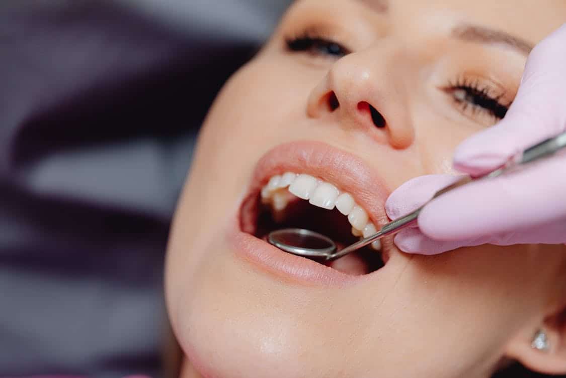
1. Oral Cancer Screening
The clinician carefully inspects the lips, tongue, cheeks, floor of the mouth, and throat for any patches, lumps, or sores that are out of the ordinary, paying close attention to texture and color changes. A visual exam is often paired with a gentle palpation and use of focused light so areas that look suspicious can be documented and tracked over time rather than left to chance.
If an abnormal spot is flagged the dental team may take photographs and recommend follow up with a specialist who can perform a biopsy if needed to determine what is going on beneath the surface. Catching malignant or premalignant tissue early greatly increases the options for treatment and reduces stress for the patient and family.
Patients sometimes feel anxious about this part of the visit yet the clinician usually explains each step in plain language and invites questions, which helps ease nerves and builds trust.
The mouth can reveal signs of systemic health issues so an exam that looks beyond the teeth serves a broader purpose, offering clues that a primary care clinician or specialist might want to explore further.
A pragmatic mindset guides the process since small findings tracked over successive visits are far easier to address than surprises found at a later stage. In short the careful eye and a steady hand at the exam make early detection a realistic goal rather than a hope.
2. Tooth Decay And Cavities
Each tooth receives a methodical look for tiny pits, soft spots, discoloration, and other signals that enamel has begun to break down, with probing and visual magnification helping to reveal areas that the patient cannot see. The clinician may use bitewing x rays to reveal decay lurking between teeth or just under existing restorations, because many early lesions hide out of plain sight until they have grown larger.
When decay is found the treatment plan aims to remove diseased tissue and restore the tooth with materials that match color and strength so chewing ability returns and sensitivity declines. Acting early often means a smaller restoration is needed which preserves natural tooth structure and lowers treatment time.
If a lesion has progressed into the nerve the dentist will explain options that range from root canal therapy to extraction and replacement, and will map out consequences and timelines for each path so the patient can weigh what fits their life.
Digital imaging and chair side photos help make explanations concrete, turning abstract risk into visible findings that feel less scary and more manageable. The goal is practical: stop destructive processes early and keep as much of the natural tooth as possible so function and comfort last. A little work now can prevent a cascade of visits later.
3. Gum Health And Periodontal Screening
The hygienist or dentist measures the space between the gum and tooth around each tooth with a probe, noting depth numbers that indicate whether tissue is healthy or whether pockets suggest active periodontal disease. Bleeding on probing, redness, recession, and looseness are all pieces of a larger puzzle that the team assembles to judge the state of the supporting structures beneath the gum.
If pockets are deep or other signs point to ongoing disease the clinician will outline nonsurgical care options such as scaling and root planing and set a follow up schedule for healing and reassessment. Preserving bone and connective tissue is a major focus because healthy gums anchor teeth and keep the smile stable for years.
Beyond procedures in the clinic the team will often coach on home care techniques that improve outcomes, showing specific brushing angles, flossing methods, and adjuncts like interdental brushes that suit the patient’s anatomy.
Lifestyle factors including tobacco use and nutrition are reviewed since they have outsize influence on healing and susceptibility to infection, and modest habit shifts can produce disproportionately large gains.
Visiting a clever dental office ensures these personalized strategies are explained clearly and monitored effectively over time.
A steady pattern of professional cleanings combined with targeted home efforts creates momentum that slows or stops progression and keeps long term costs lower. The exam thus acts as both diagnostic check and a launch point for better daily habits that protect the investment.
4. Bite And Jaw Alignment
The dentist observes how upper and lower teeth contact each other in various jaw positions and watches for uneven wear facets, chips, or cracks that suggest grinding, clenching, or a misaligned bite that places stress on specific teeth.
The temporomandibular joint is evaluated for smooth opening and closing, with attention to clicks, catches, or pain that can point to muscular imbalance or joint dysfunction that affects chewing and comfort.
When a functional problem is identified the clinician will present options ranging from conservative splint therapy and home exercises to occlusal adjustments or collaboration with orthodontics if tooth position is part of the issue. Restoring a balanced bite often reduces headaches and neck tension and protects teeth from accelerated breakdown.
For growing patients the team tracks tooth eruption and jaw development so early interventions can guide alignment before patterns harden, while for adults the focus shifts to restoring lost vertical height or managing wear that has built up over many years.
Nighttime guards and behavioral strategies to reduce clenching are common low risk steps that can stabilize symptoms while longer term plans are formed. In cases that require more extensive correction the dentist coordinates with specialists so care flows smoothly and goals remain clear. The objective is practical comfort and durable function rather than cosmetic perfection alone.
5. X Rays And Imaging Review
Radiographs reveal the hidden landscape beneath tooth enamel, showing bone support, roots, impacted teeth, and areas of decay that escape visual inspection, and modern digital systems let the clinician enhance and compare images quickly.
These images complement the hands on exam, offering evidence that explains findings and informing whether monitoring, conservative care, or more invasive steps are warranted. When infection or bone loss appears the image helps map its extent and shape the treatment sequence so interventions hit the problem where it lives rather than guessing.
Clear imaging increases confidence for both clinician and patient and reduces the chance of surprises in the treatment chair.
For procedures that require detailed planning, such as implant placement or surgical extraction, three dimensional scans provide added precision by showing nerve pathways and bone contours in real space so risks are minimized and outcomes become more predictable.
Patient concerns about exposure are addressed with data about low dose protocols and the trade off between a slightly higher dose and the value of targeted, accurate care planning. Visuals also support a collaborative conversation where the person in the chair sees what the team sees, making choices feel more informed and less abstract. Good imaging is a tool that turns uncertainty into a clear map for action.


